
Bone Disorder Comparison Tool
Cell Imbalance: Excessive osteoclast activity followed by disorganized osteoblast activity
Age Group: Typically affects individuals aged 50-80 years
Common Sites: Pelvis, spine, skull, femur
Cell Imbalance: Increased bone resorption with reduced formation
Age Group: Post-menopausal women and elderly (>65 years)
Common Sites: Vertebrae, hip, wrist
Cell Imbalance: Cartilage degeneration leading to subchondral bone changes
Age Group: 40+ years, more common in older adults
Common Sites: Knees, hips, hands
Cell Imbalance: Replacement of normal bone with fibrous tissue
Age Group: Often diagnosed in teens to 20s
Common Sites: Long bones, ribs, craniofacial bones
| Feature | Paget's Disease | Osteoporosis | Osteoarthritis | Fibrous Dysplasia |
|---|---|---|---|---|
| Radiographic Hallmark | Mixed lytic-sclerotic lesions, "cotton-wool" skull | Trabecular thinning, cortical porosity | Joint space narrowing, osteophytes | Ground-glass appearance, well-defined borders |
| Blood Marker | Elevated ALP (often 2-3× normal) | Normal ALP; low BMD on DXA | Normal ALP; inflammatory markers may rise | Usually normal ALP unless active lesion |
| First-Line Treatment | Bisphosphonates (zoledronic acid) | Calcium + Vitamin D, bisphosphonates | Analgesics, physical therapy | Surgical resection of symptomatic lesions |
- Check serum alkaline phosphatase; elevated values warrant imaging
- Order a bone scan if Paget's is suspected; it visualizes the whole skeleton
- Use DXA to rule out osteoporosis; interpret results cautiously if Paget's lesions are present
- Consider bisphosphonate therapy-IV for Paget's, oral for osteoporosis
- Screen for vitamin D deficiency; correct it before bisphosphonate treatment
When doctors spot abnormal bone growth or pain, they often wonder whether it’s Paget's disease, osteoporosis, osteoarthritis, or something else. Understanding how these conditions overlap-and where they diverge-helps patients get the right tests, treatment, and peace of mind.
What is Paget's Disease?
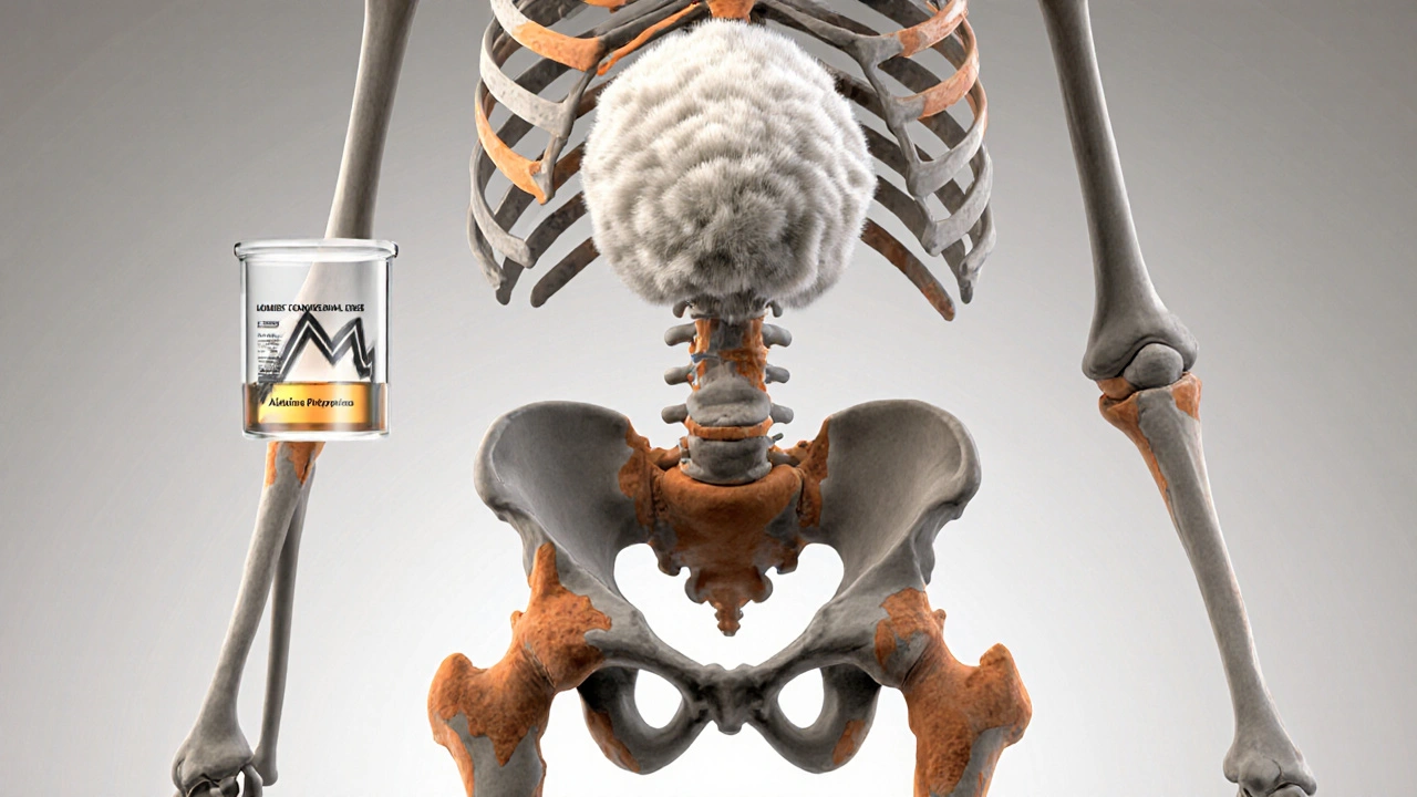
Paget's disease is a chronic disorder that disrupts the normal cycle of bone remodeling. Instead of a balanced dance between bone‑building cells (osteoblasts) and bone‑breaking cells (osteoclasts), the disease triggers osteoclasts to work overtime, creating weak, enlarged bone that later fills in with irregular, dense tissue.
Typical signs include bone pain, noticeable enlargement of the skull or long bones, and in some cases hearing loss when the skull is involved. Blood tests often reveal elevated alkaline phosphatase (ALP), while X‑rays show a classic “cotton‑wool” appearance.
Bone Remodeling: The Core Process Behind All These Disorders
All the bone disorders we’ll discuss share a common foundation: the remodeling cycle. Osteoclast cells dissolve old bone, and osteoblast cells lay down new matrix. Hormones like parathyroid hormone, vitamin D, and calcium levels fine‑tune this balance.
When the balance tips-whether because osteoclasts become overactive (Paget's), underactive (osteopetrosis), or simply age‑related-bone density, strength, and shape suffer.
Other Common Bone Disorders That Share Pathways
Osteoporosis is the opposite problem: bone resorption outpaces formation, leading to porous, fragile skeletons. It’s driven largely by hormonal shifts post‑menopause, low calcium intake, and sedentary lifestyles.
Osteoarthritis isn’t a remodeling disorder per se, but the wear‑and‑tear of cartilage changes joint loading, prompting subchondral bone thickening and osteophyte formation.
Fibrous dysplasia is a genetic mosaic condition where normal bone is replaced by fibrous tissue, producing painless swellings that can fracture easily.
Despite different triggers, these diseases intersect at the level of calcium metabolism, ALP elevation, and imaging findings, which can confuse diagnosis.
Side‑by‑Side Clinical Comparison
| Feature | Paget's Disease | Osteoporosis | Osteoarthritis | Fibrous Dysplasia |
|---|---|---|---|---|
| Primary Cell Imbalance | Excessive osteoclast activity, later disorganized osteoblast activity | Increased resorption, reduced formation | Cartilage degeneration, reactive subchondral bone | Replacement of normal bone with fibrous tissue |
| Typical Age | 50‑80 years | Post‑menopausal women, >65 years | 40+ years, more common in older adults | Often diagnosed in teens‑20s |
| Common Sites | Pelvis, spine, skull, femur | Vertebrae, hip, wrist | Knees, hips, hands | Long bones, ribs, craniofacial bones |
| Radiographic Hallmark | Mixed lytic‑sclerotic lesions, "cotton‑wool" skull | Trabecular thinning, cortical porosity | Joint space narrowing, osteophytes | Ground‑glass appearance, well‑defined borders |
| Blood Marker | Elevated ALP (often 2‑3× normal) | Normal ALP; low BMD on DXA | Normal ALP; inflammatory markers may rise | Usually normal ALP unless active lesion |
| First‑Line Treatment | Bisphosphonates (e.g., zoledronic acid) | Calcium + Vitamin D, bisphosphonates, lifestyle | Analgesics, physical therapy, joint replacement if severe | Surgical resection of symptomatic lesions |
Diagnosing Overlap - When Tests Blur the Lines
Because several disorders raise alkaline phosphatase and alter bone density, doctors usually start with a detailed history and physical exam, then order a cascade of investigations:
- Blood work: ALP, calcium, phosphate, vitamin D, and specific markers like procollagen type1N‑terminal propeptide (P1NP) for bone formation.
- Dual‑energy X‑ray absorptiometry (DXA): Shows low BMD in osteoporosis but can be misleading if Paget's lesions are present, as they artificially raise density.
- Plain radiographs: Provide the first visual clue-cotton‑wool skull for Paget's, trabecular thinning for osteoporosis, joint space narrowing for osteoarthritis.
- Bone scintigraphy (bone scan): Highly sensitive for Paget's; the affected area lights up intensely.
- CT / MRI: Used when structural details matter-e.g., to assess skull involvement or differentiate a fibrous dysplasia lesion from a metastatic deposit.
Combining these tools lets clinicians tease apart conditions that otherwise look alike.
Shared and Distinct Treatment Pathways
Bisphosphonates-drugs that inhibit osteoclasts-are the mainstay for both Paget's disease and osteoporosis. However, the dosing differs: a single intravenous zoledronic acid infusion often normalizes ALP in Paget's within weeks, whereas osteoporosis may need oral alendronate weekly for years.
Calcitonin, once a go‑to for Paget's, has largely been replaced by newer bisphosphonates but still appears in guidelines for patients who cannot tolerate them.
Osteoarthritis treatment rarely targets bone remodeling; it focuses on pain control (acetaminophen, NSAIDs), joint preservation (physical therapy), and surgical replacement when cartilage loss is severe.
Fibrous dysplasia management is mostly surgical, though bisphosphonates can reduce bone pain in active lesions.
Genetic and Environmental Links
Research shows that mutations in the SQSTM1 gene increase susceptibility to Paget's disease by enhancing osteoclast activity. Interestingly, some of the same signaling pathways (RANK/RANKL/OPG) are implicated in osteoporosis, suggesting that a family history of one bone disease may hint at heightened risk for another.
Environmental factors-like viral infections (paramyxoviruses) once theorized to trigger Paget's-also influence immune-mediated bone loss seen in rheumatoid arthritis, blurring the lines further.
Practical Checklist for Patients and Clinicians
- Ask about bone pain, especially in the pelvis, spine, or skull.
- Check serum alkaline phosphatase; a high value warrants imaging.
- Order a bone scan if Paget's is suspected; it visualizes the whole skeleton.
- Use DXA to rule out osteoporosis; interpret results cautiously if Paget's lesions are present.
- Consider bisphosphonate therapy-IV for Paget's, oral for osteoporosis-based on diagnosis.
- Screen for vitamin D deficiency; correct it before or alongside bisphosphonate treatment.
- Refer to orthopedics if deformities or fractures develop.
- Monitor ALP every 3-6months after starting treatment for Paget's.
Key Takeaway
Paget's disease uniquely blends excessive bone breakdown with chaotic rebuilding, but its clinical and laboratory fingerprints often overlap with osteoporosis, osteoarthritis, and fibrous dysplasia. Recognizing the subtle differences-especially through targeted imaging and blood work-ensures patients receive the right therapy and avoid unnecessary procedures.

Frequently Asked Questions
Can Paget's disease cause fractures?
Yes. The disorganized bone can become thick yet brittle, making it prone to fractures, especially in the femur and pelvis.
How is Paget's disease different from osteoporosis on an X‑ray?
Paget's shows mixed lytic and sclerotic patches, often described as "cotton‑wool" appearance, while osteoporosis displays generalized bone thinning without focal lesions.
Are bisphosphonates safe for both Paget's and osteoporosis?
Generally, yes. They inhibit osteoclasts, which helps both conditions. However, dosing schedules differ, and patients should be screened for kidney function before IV administration.
Is there a genetic test for Paget's disease?
Testing for SQSTM1 mutations can confirm a hereditary form, but most cases are diagnosed clinically and with imaging.
Can lifestyle changes prevent Paget's disease?
Since the exact cause is unknown, no specific lifestyle tweak guarantees prevention. Maintaining adequate calcium and vitamin D, however, supports overall bone health and may lower the risk of related disorders like osteoporosis.


Comments (10)